
Best Biology Specimens for Classroom Study & Dissection
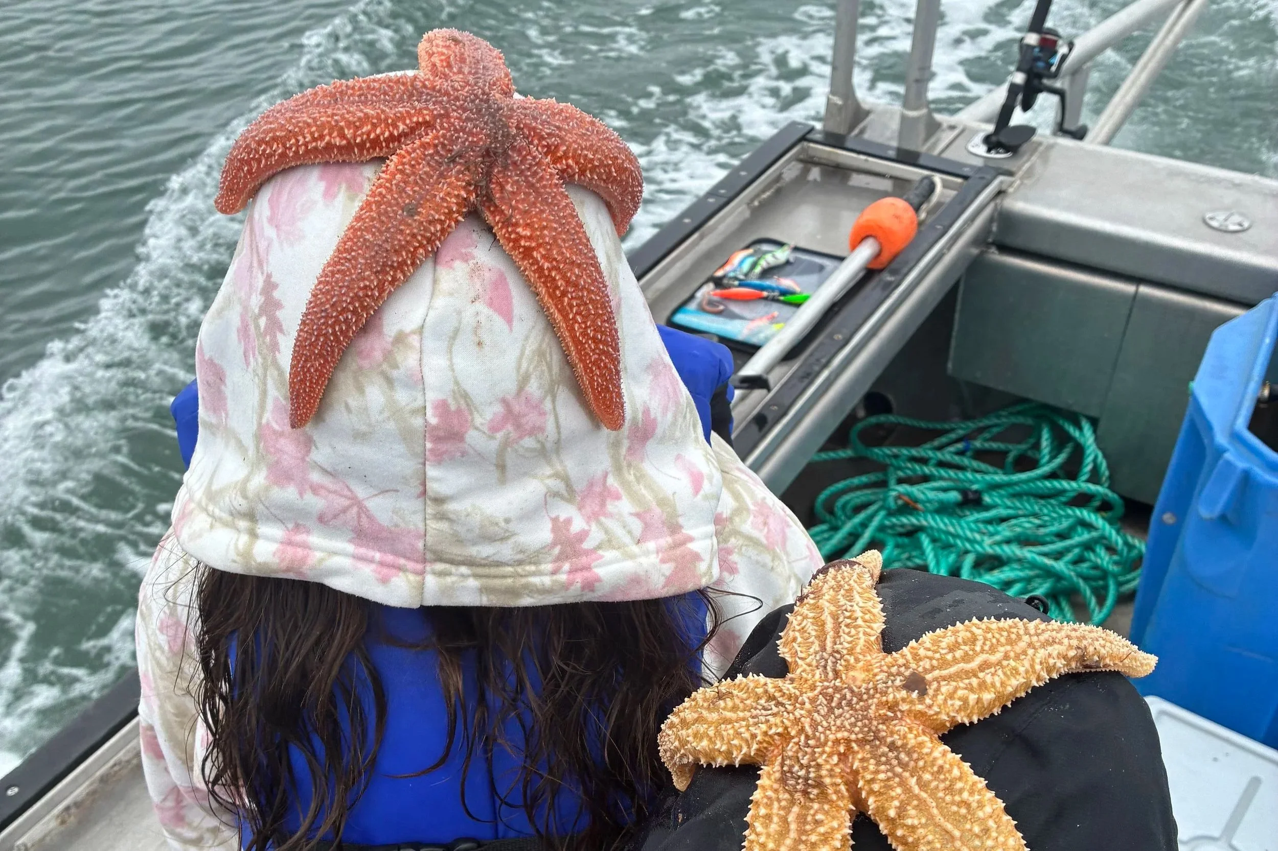
After more than 25 years of supplying biological specimens to classrooms and labs, I've learned that there really isn't one "best" specimen to dissect. It depends on what you want your students to see.
A yellow perch and a spiny dogfish are both fish, but put them side by side and suddenly you have a lesson in two very different approaches to being a vertebrate. A squid and a freshwater mussel hardly look like relatives at all, yet both belong to the phylum Mollusca. And a sea star? Well, that takes the familiar animal body plan and more or less throws it out the window.
That's what makes working with real biology specimens so valuable.
When I taught biology, I always found that the more students could actually see, touch, compare, and investigate, the easier it was for them to understand concepts that can seem pretty abstract on a textbook page.
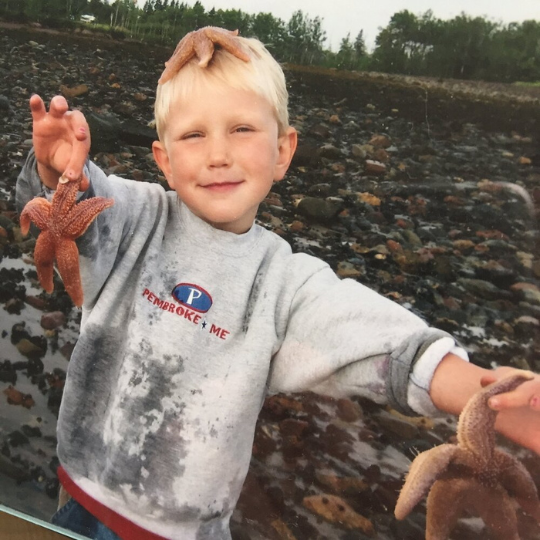
My Son and I tidepooling in 2004
So rather than simply giving you a list of dissection specimens, here are some of the animals I'd choose depending on what you're trying to teach.
Start With a Classic: Yellow Perch
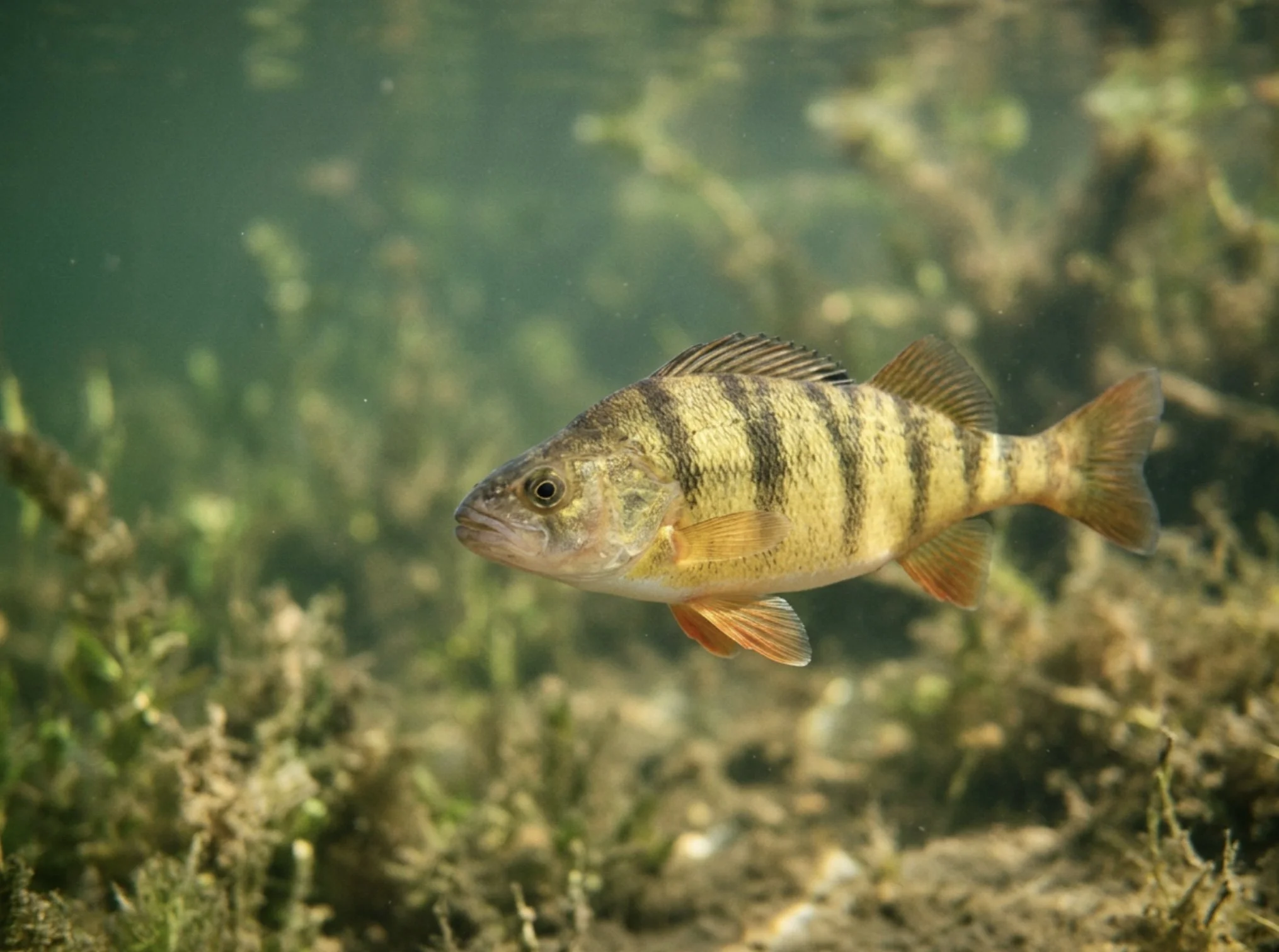
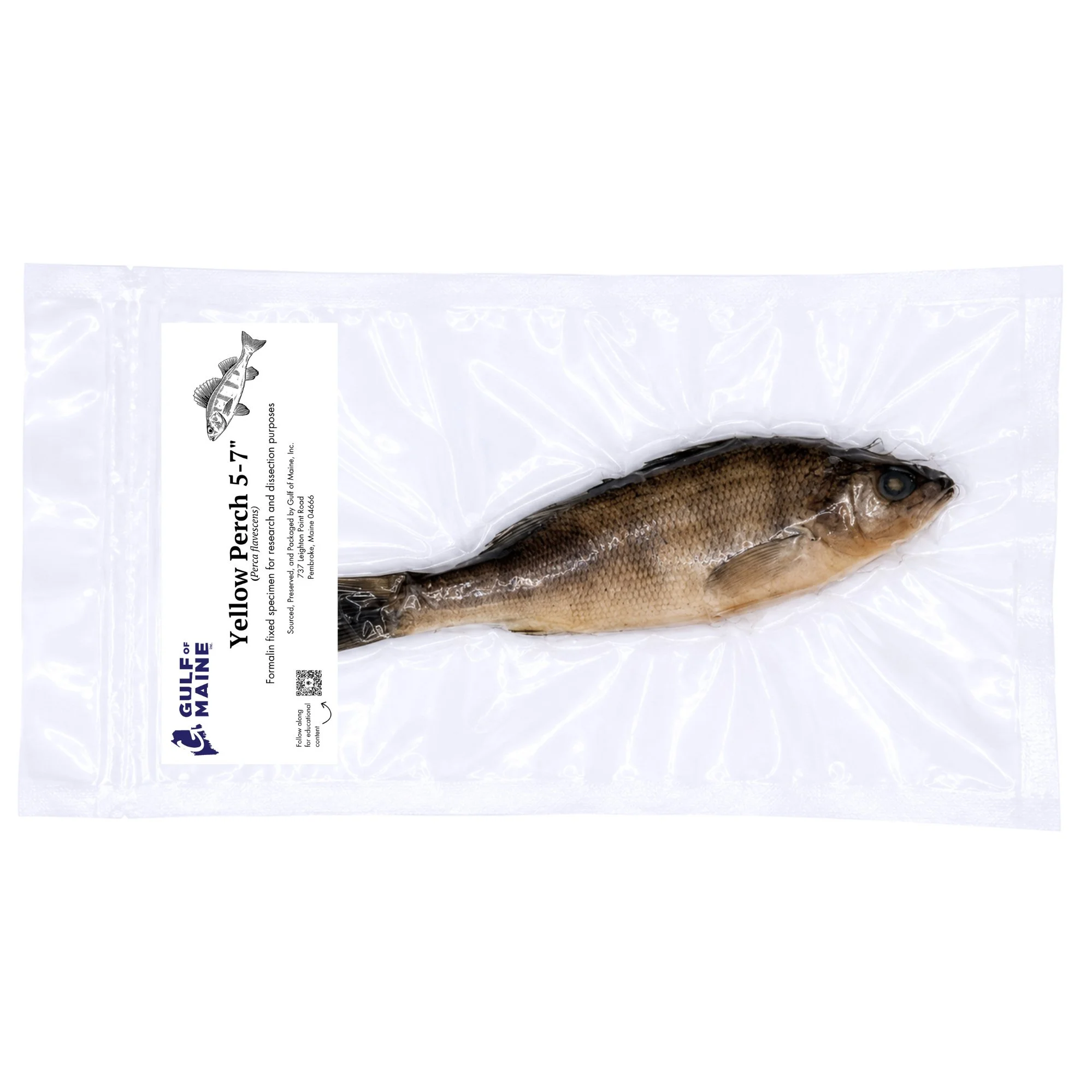
There's a reason yellow perch (Perca flavescens) have been showing up on biology lab tables for generations.
They're an excellent introduction to the anatomy of a bony fish, and there's plenty to investigate before you ever pick up a scalpel.
Externally, students can identify the fins, scales, lateral line, mouth, eyes, and operculum. From there, a dissection reveals the gills, heart, liver, stomach, intestine, swim bladder, reproductive organs, and other internal structures.
The swim bladder is particularly useful for connecting structure with function. Instead of simply memorizing the name of another organ, students can think about a practical problem faced by a fish: how do you maintain your position in the water without constantly swimming upward or sinking to the bottom?
That's when anatomy starts becoming biology rather than a vocabulary exercise.
Yellow perch are also useful because they give students a familiar vertebrate body plan that can later be compared with something considerably different.
Which brings us to a shark.
Now Take Away the Bones: Spiny Dogfish
The spiny dogfish (Squalus acanthias) is another classic dissection specimen, but it gives students a very different look at fish anatomy.
Dogfish are sharks and belong to the cartilaginous fishes. Rather than having a skeleton made primarily of bone like a yellow perch, their skeleton is made of cartilage.
Internally, students can examine structures including the gills, heart, liver, stomach, intestine, and reproductive organs. Externally, there are also plenty of adaptations to investigate, including the fins, gill slits, spiracles, lateral line, and—true to the name—the spines located in front of the dorsal fins.
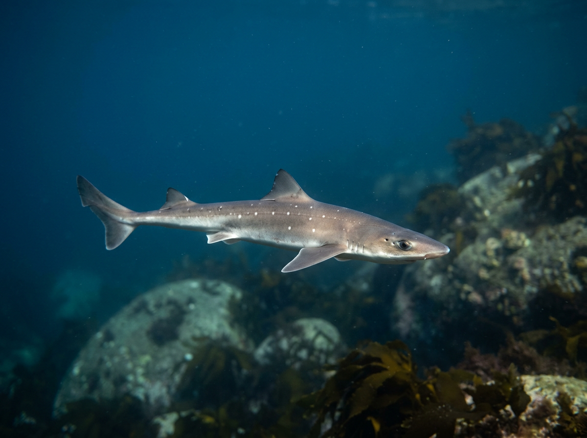
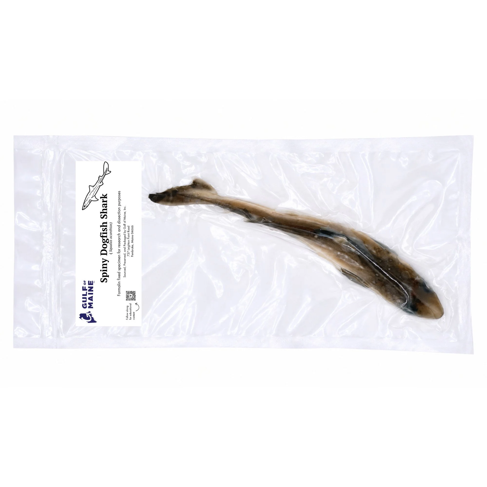
Yellow Perch vs. Spiny Dogfish
If you have the opportunity, these are two specimens I'd strongly recommend putting side by side.
Both are fishes. Both are vertebrates. Both have to solve many of the same basic problems: breathe underwater, find food, move efficiently, reproduce, and keep themselves at the appropriate depth.
But they don't solve all of those problems in the same way.
Students can compare the bony skeleton of a perch with the cartilaginous skeleton of a dogfish, an operculum with exposed gill slits, and the swim bladder of a perch with the very different buoyancy adaptations of a shark.
That's the real value of comparative anatomy. Instead of asking only, "What is this structure called?" you can start asking, "Why is it different?"
Squid: When the Body Plan Gets Interesting
If your students have spent most of their time studying vertebrates, put a squid in front of them.
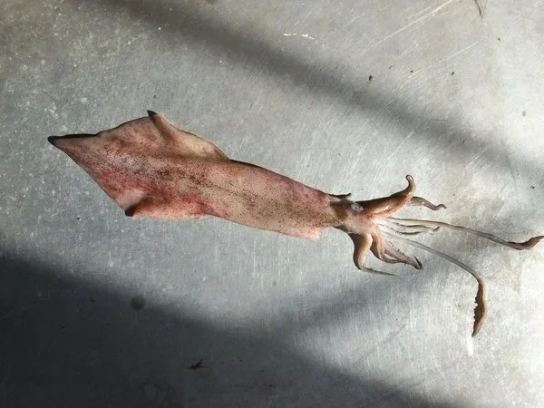
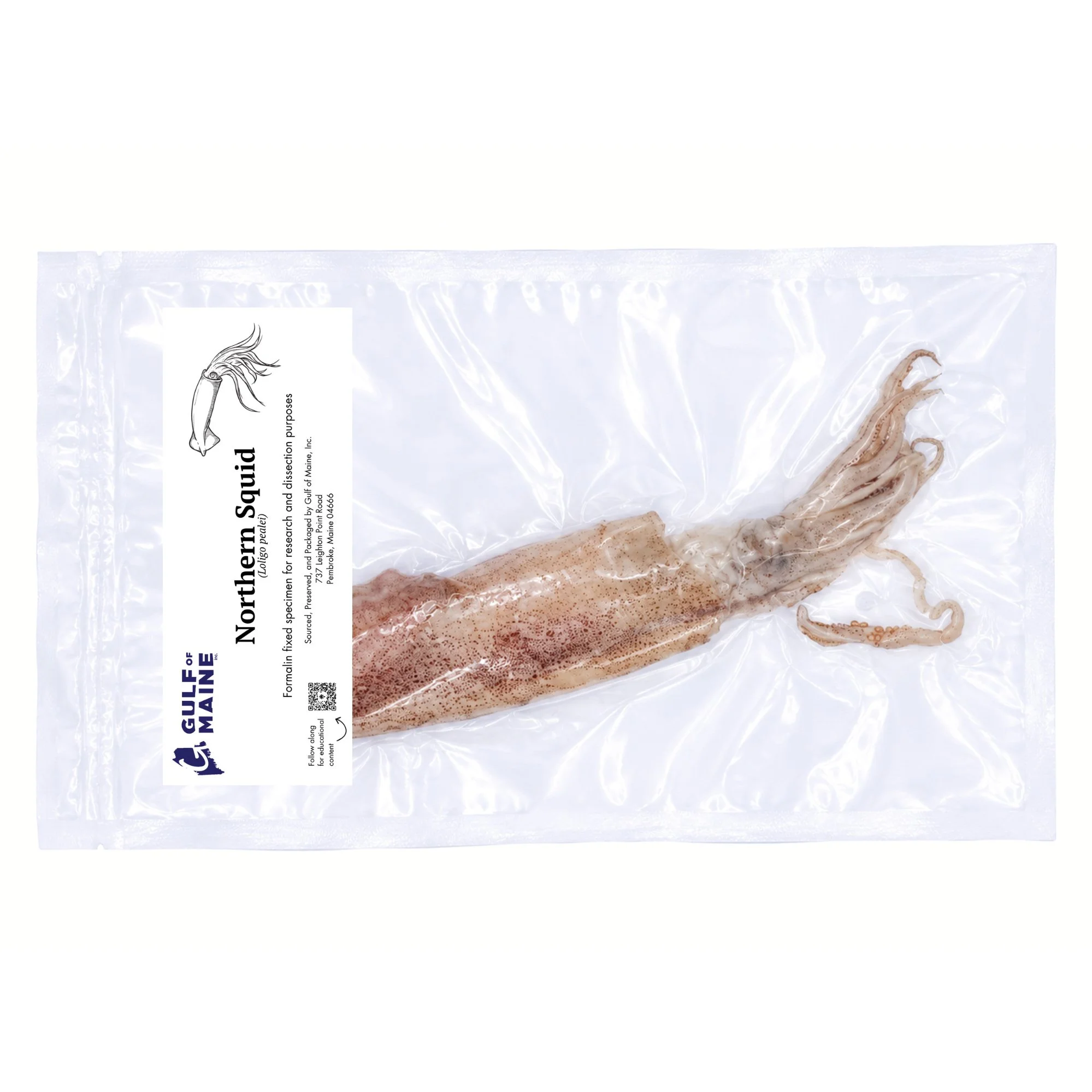
Squid are cephalopod mollusks, and their anatomy looks remarkably different from that of a fish. The mantle forms the main body, the foot characteristic of mollusks has been modified into arms, tentacles, and the siphon, and an internal structure called the pen is all that remains of the shell found in many of their mollusk relatives.
A squid dissection gives students a chance to examine the mantle, arms, tentacles, suckers, siphon, gills, ink sac, beak, digestive organs, and reproductive structures.
They're also great animals for discussing how anatomy relates to behavior.
The siphon allows a squid to move using jet propulsion. The arms and tentacles are equipped with suckers for handling and capturing prey. Hidden where all those arms meet is a hard beak used to bite food.
And yes, the ink sac tends to get everyone's attention.
There's a lot going on inside a squid, which makes it one of my favorite specimens for getting students to think about how a very different animal body plan accomplishes familiar tasks.
Freshwater Mussels: Yes, They're Related to the Squid
Now put a freshwater mussel next to that squid.
At first glance, asking students what the two animals have in common might earn you a few puzzled looks. That's exactly why it's a useful comparison.
Both belong to the phylum Mollusca.
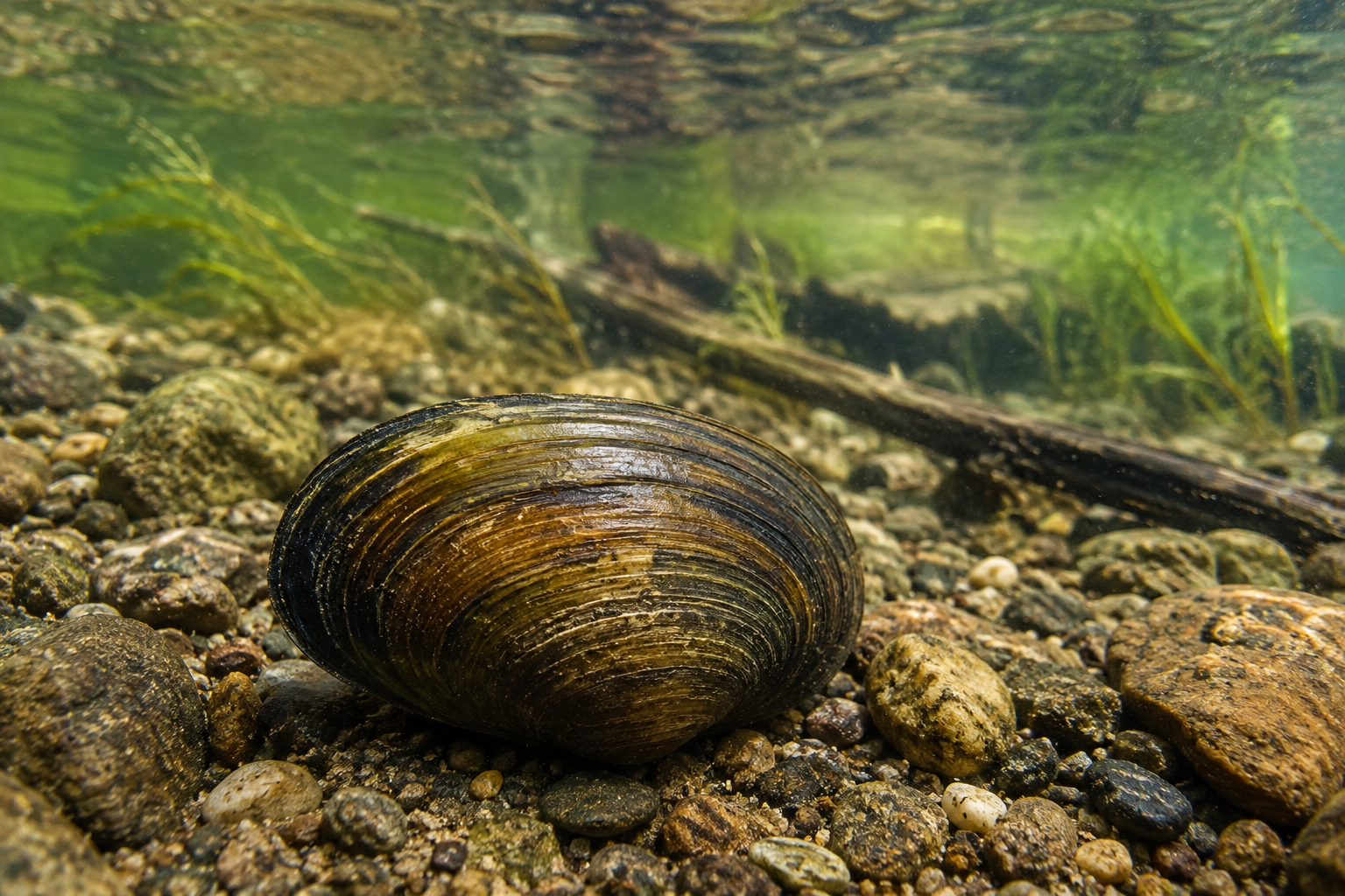
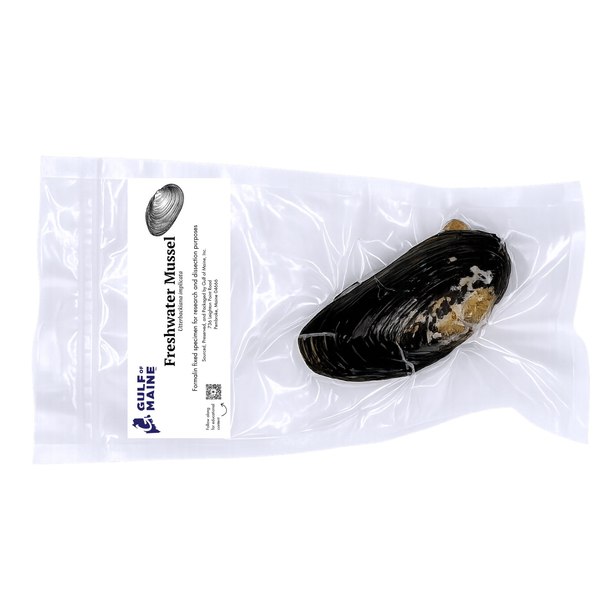
Freshwater mussels have a very different lifestyle and anatomy from an active, predatory cephalopod. Students can examine the shell and hinge, mantle, gills, adductor muscles, muscular foot, and digestive structures while looking at how the animal is adapted for life as a bivalve.
The gills are especially interesting because they aren't simply respiratory structures. They also play an important role in filter feeding.
Two Mollusks, Two Very Different Animals
Comparing a freshwater mussel and squid is a good reminder that belonging to the same phylum doesn't mean two animals have to look much alike.
One is protected by two shells and spends its life associated with the bottom. The other swims through the water using jet propulsion and captures prey with its arms and tentacles.
Yet underneath those differences are characteristics that place both animals within Mollusca.
This is one of those comparisons that makes classification much more interesting than memorizing a list of phyla.
Sea Stars: Five Arms and a Completely Different Way of Doing Things
Sea stars are familiar animals, but their anatomy is anything but ordinary.
Adult sea stars have pentaradial symmetry rather than the bilateral symmetry we see in ourselves, fishes, squid, and many other animals. Turn one over and you'll find rows of tube feet extending down the underside of the arms.
Those tube feet are part of the sea star's water vascular system, a hydraulic system involved in movement, feeding, and attachment.
During a sea star dissection, students can examine the stomachs, digestive glands, gonads, tube feet, ambulacral grooves, and components of the water vascular system.
The digestive system is another good example of why sea stars are worth studying. A sea star can extend part of its stomach outside of its body to begin digesting prey externally. If you've ever wondered how an animal with no teeth manages to eat a mussel, that's part of the answer.
We've already put together a complete Northern Sea Star Dissection Guide for anyone who wants to go through the anatomy in more detail.
Then Put a Sea Urchin Beside It
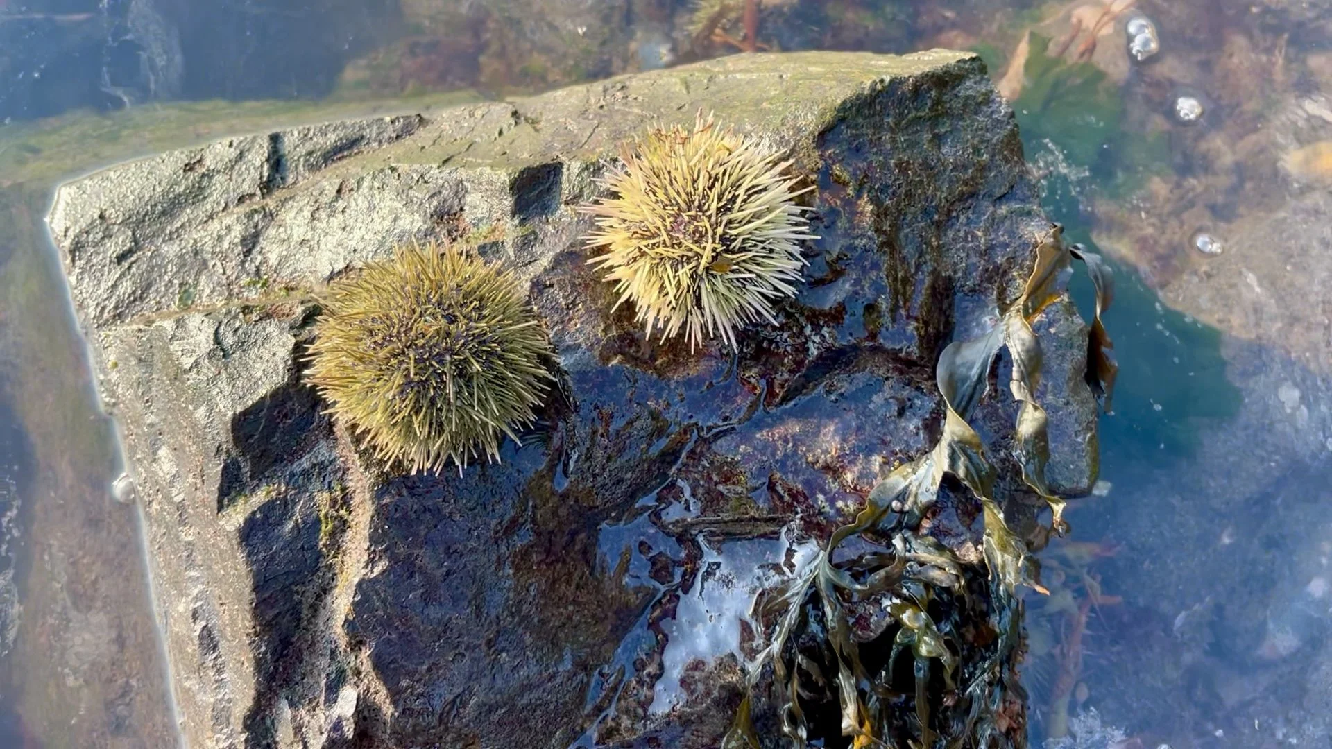
A sea urchin doesn't look much like a sea star.
One has arms. The other looks like a spiny ball.
Yet both belong to the phylum Echinodermata, and looking closely reveals some of the characteristics they share.
Students can compare their five-part organization, tube feet, water vascular systems, external structures, feeding adaptations, and methods of locomotion.
A sea urchin's hard internal skeleton, called a test, gives it a very different appearance from a sea star, but the underlying echinoderm body plan is still there.
This is another pairing I like because students have to look beyond an animal's overall appearance to figure out what actually connects the two.
Crayfish: An Arthropod You Can Really Examine
Crayfish are another classroom classic, and for good reason.
Their hard exoskeleton and jointed appendages make many anatomical features easy to see before the specimen is even opened.
Students can identify the cephalothorax and abdomen, antennae, walking legs, swimmerets, mouthparts, and other external structures. Internally, they can examine the gills, digestive system, reproductive structures, muscles, and other organs.
They're particularly useful for teaching the arthropod body plan: segmentation, a protective exoskeleton, and specialized jointed appendages.
A crayfish also provides a nice contrast to many of the other specimens in this guide. Compare one with a sandworm and both show segmentation—but in very different ways. Compare it with a squid and you have two invertebrates with entirely different systems of support and movement.
Sometimes the differences are the lesson.
Don't Overlook the Worms
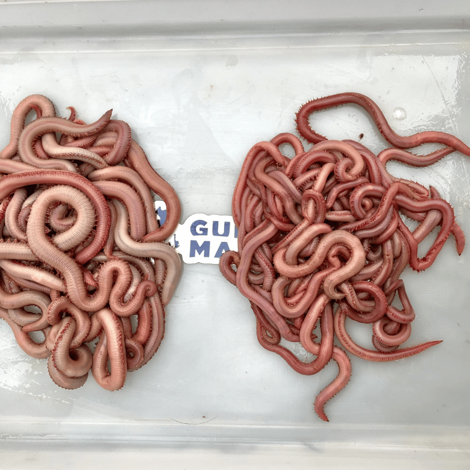
Sandworms and bloodworms aren't usually the specimens that get students excited when you first announce a dissection.
Give them a chance.
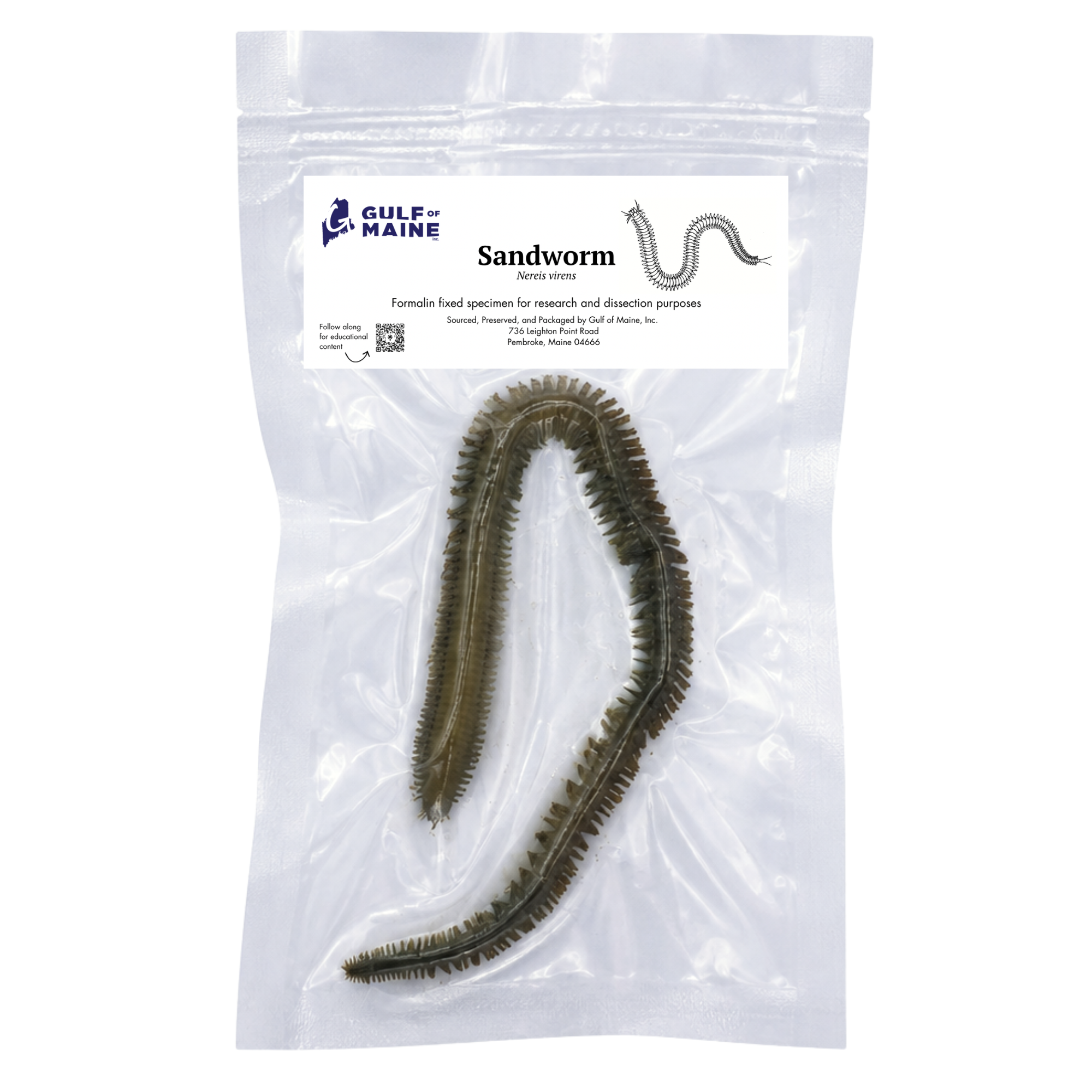
Sandworms and bloodworms are polychaete annelids, and their segmented bodies provide a very clear example of the annelid body plan.
Along the body, students can examine repeated segments as well as structures such as parapodia and setae. These structures are involved in locomotion and help the worms move through or across marine sediments.
They're also a good opportunity to connect anatomy with ecology.
Marine worms may not get the attention that sharks, sea stars, and squid do, but they're an important part of coastal food webs. They live within the sediment, process organic material, and serve as prey for fishes, crustaceans, and shorebirds.
Sometimes one of the least glamorous animals on the lab table has one of the biggest ecological jobs.

Sea Squirts: The Chordate That Doesn't Look the Part
If you really want to challenge students' assumptions about animal classification, give them a sea squirt.
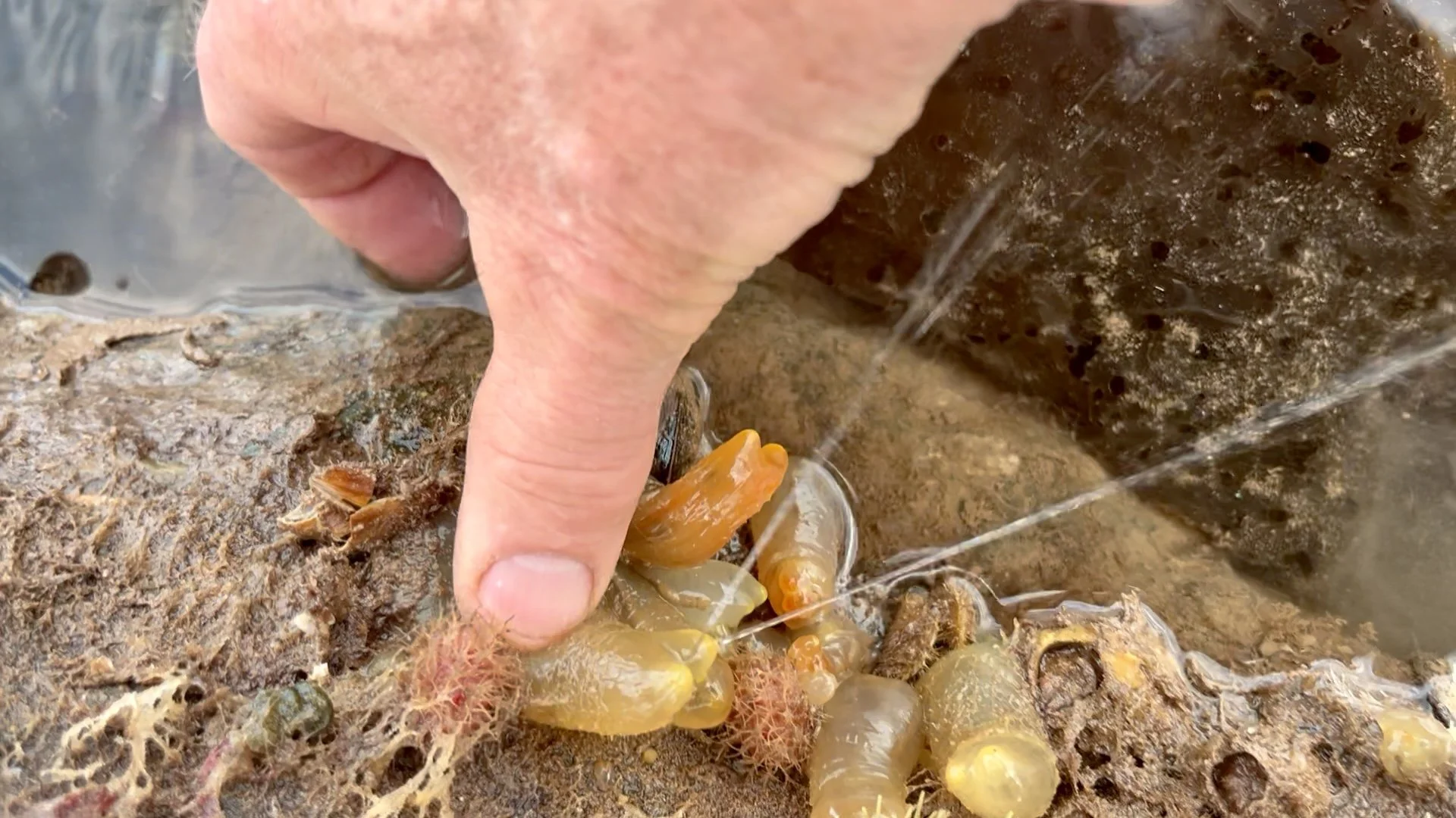
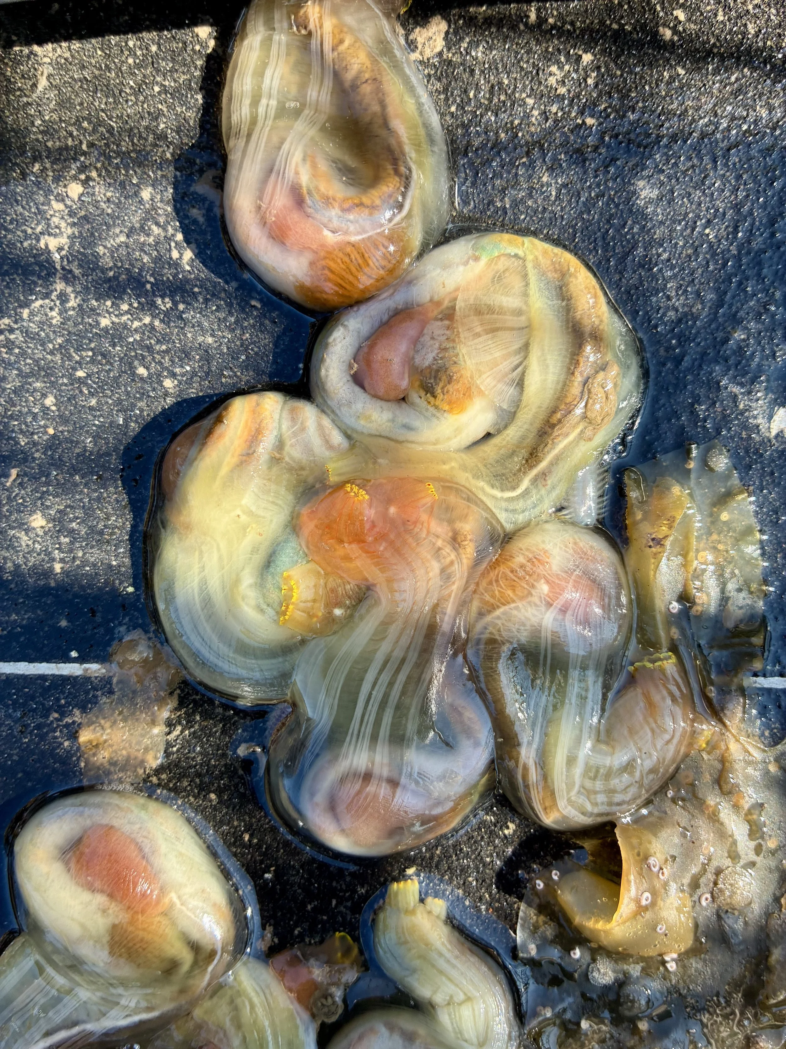
Sea squirts, or tunicates, don't look much like fishes—or us—but they're members of the phylum Chordata.
An adult sea squirt such as a sea vase (Ciona spp.) is essentially a stationary filter feeder. Water enters through one siphon, passes through a large pharyngeal basket where suspended particles are captured, and exits through another siphon.
The really interesting part comes when you look at the larval stage.
Tunicate larvae possess chordate characteristics that are much easier to recognize, including a notochord and dorsal nerve cord. Those structures are lost or greatly reduced as the animal transforms into the adult form.
So if students are wondering why the strange bag-shaped animal sitting on the lab table belongs anywhere near a fish on the tree of life, they're asking exactly the right question.
So, Which Biology Specimen Should You Choose?
There really isn't a single answer.
If you're teaching bony fish anatomy, start with a yellow perch.
For cartilaginous fishes and shark anatomy, use a spiny dogfish. Better yet, compare the two.
For mollusks, squid are excellent dissection specimens, while pairing a squid with a freshwater mussel demonstrates just how diverse Mollusca can be.
For echinoderms, sea stars provide a good introduction to radial symmetry and the water vascular system. Add a sea urchin and students can look for the characteristics shared by two animals that, at first glance, seem completely different.
Crayfish provide a clear look at the arthropod body plan, while marine worms demonstrate annelid segmentation.
And if you're discussing Chordata, a sea squirt can make students reconsider what they think a chordate is supposed to look like.
The important thing isn't necessarily choosing the animal with the most structures to label. It's choosing a specimen—or combination of specimens—that helps answer the biological question you're teaching.
Preserved Specimens Aren't Just for Dissection
It's worth pointing out that you don't necessarily have to dissect every preserved specimen you bring into the classroom.
Real specimens can be useful for studying external anatomy, taxonomy, adaptation, biodiversity, species identification, ecology, comparative anatomy, and form and function.
Sometimes simply placing several organisms beside one another is enough to start a good lesson.
Give students a yellow perch, dogfish, squid, sea star, crayfish, and mussel and ask them to sort the animals based on characteristics they can observe. Then ask them to explain their groupings.
You'll probably get some interesting answers.
From there, you can introduce taxonomy and see how their observations compare with the relationships biologists use to classify the animals.
For educators who are new to preserved material, we've also put together a Preserved Marine Specimens for Dissection guide that covers preservation, handling, storage, and classroom use.
Building a Comparative Biology Lab
If I were putting together a collection specifically for comparative anatomy, I'd try to represent several different animal groups rather than choosing a number of specimens that all teach essentially the same lesson.
For example:
Yellow Perch — Chordata, bony fish
Spiny Dogfish — Chordata, cartilaginous fish
Squid — Mollusca, cephalopod
Freshwater Mussel — Mollusca, bivalve
Sea Star — Echinodermata
Crayfish — Arthropoda
Sandworm — Annelida
Now you've got animals with different types of symmetry, support structures, methods of locomotion, feeding strategies, respiratory structures, and organ systems.
Students can look at each animal individually, but they can also start asking larger questions.
Why does this animal have an exoskeleton while another has an internal skeleton? Why does one animal have gills protected beneath an operculum while another has exposed gill slits? How can a squid and mussel possibly belong to the same phylum? Why does a sea star have radial symmetry when so many other animals are bilateral?
Those are the questions that make a collection of preserved specimens more than a series of dissections.
Real Specimens Are Supposed to Vary
One final point that can be easy to forget when working from textbooks and diagrams: real animals aren't identical.
The specimens we work with naturally vary in size, shape, color, sex, condition, and sometimes anatomy. A structure may not look exactly like the perfectly colored illustration in a dissection manual.
That's biology.
Learning to observe what's actually in front of you rather than searching for an exact copy of a diagram is an important scientific skill in its own right.
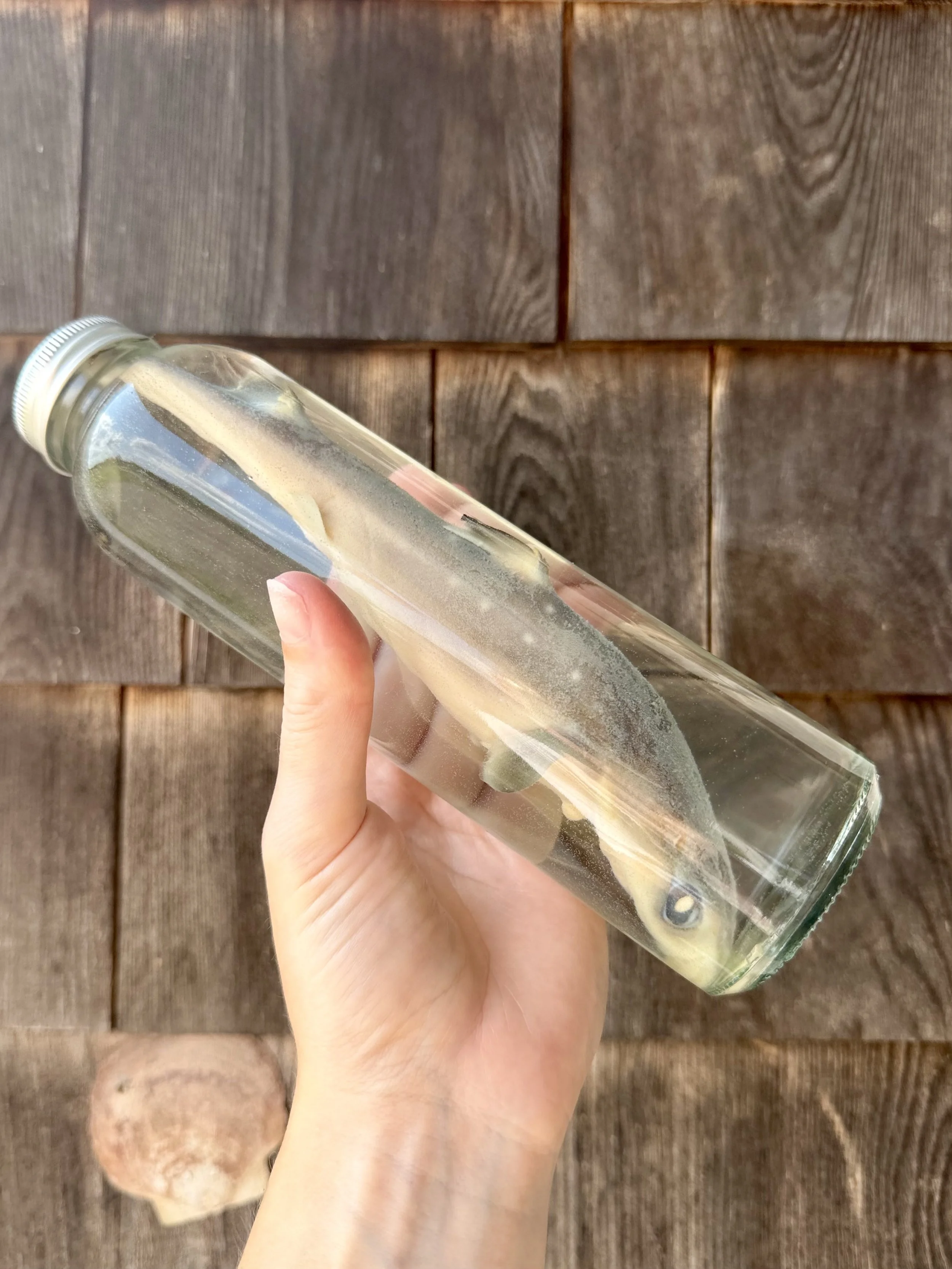
Preserved Spiny Dogfish Shark from Gulf of Maine, Inc.
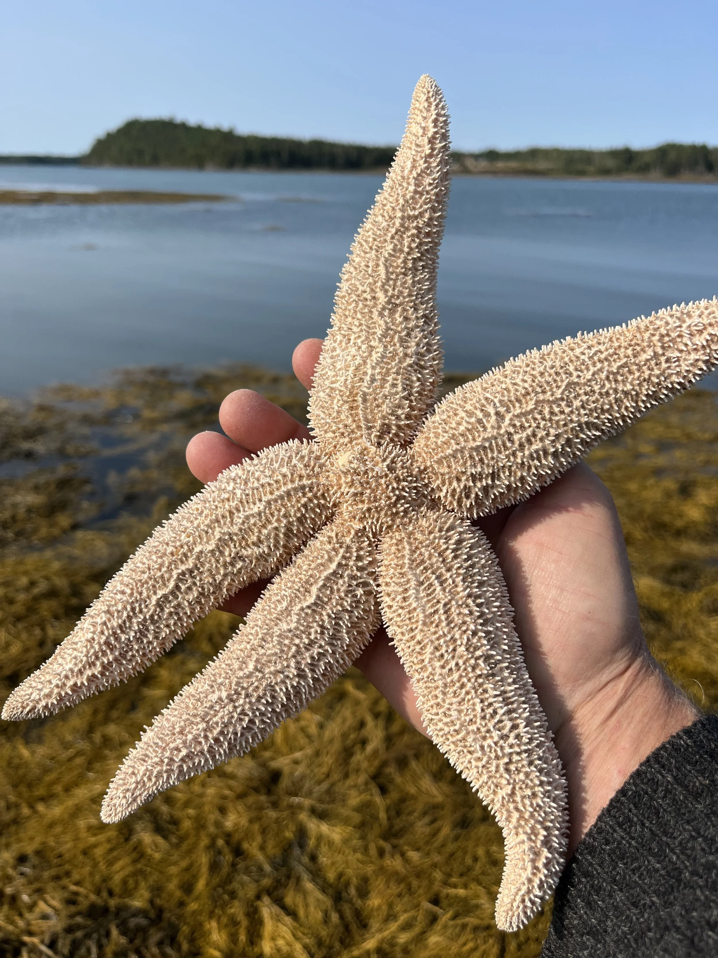
Dried Northern Sea Star from Gulf of Maine, Inc.
Biology Specimens from Gulf of Maine, Inc.
I've spent much of my career teaching people about aquatic life, and Gulf of Maine, Inc. grew out of that same belief in hands-on learning.
Since 2000, we've supplied biological specimens and aquatic life to schools, universities, laboratories, researchers, homeschoolers, and other educators.
Our preserved collection includes familiar classroom dissection specimens such as yellow perch, spiny dogfish, squid, sea stars, crayfish, and freshwater mussels, along with some less familiar animals that can bring something different to a biology lesson.
Whether you're planning one dissection or building an entire comparative anatomy unit, the goal is the same: get students looking closely, asking questions, and figuring out why an animal is built the way it is.
That's when things get interesting.
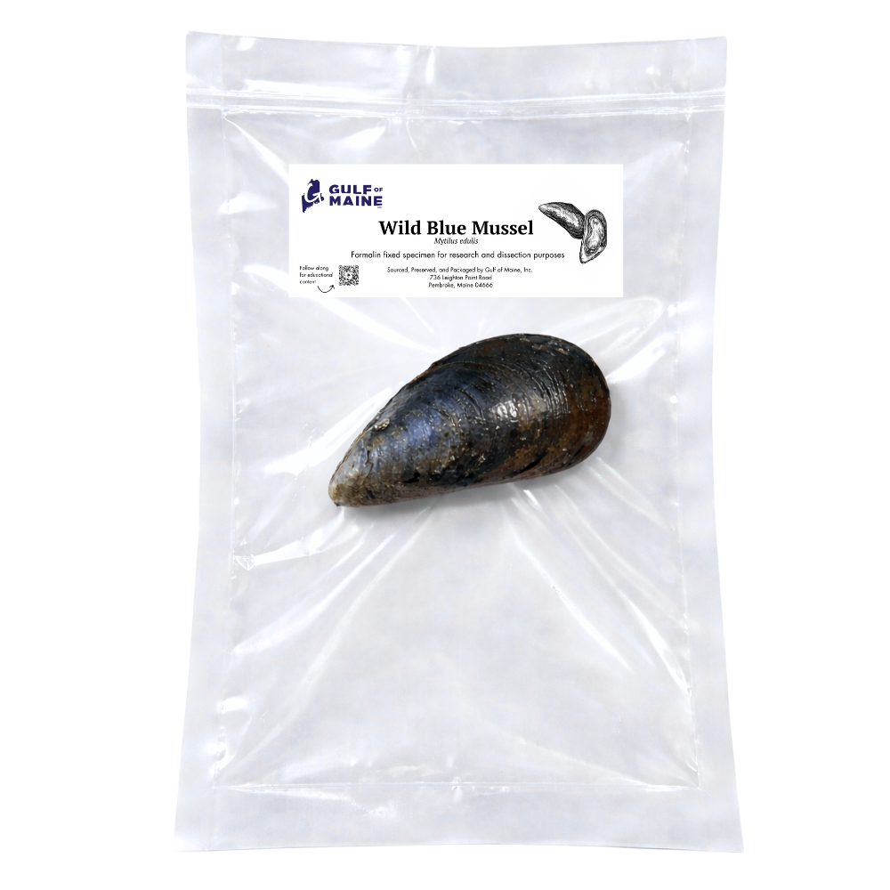
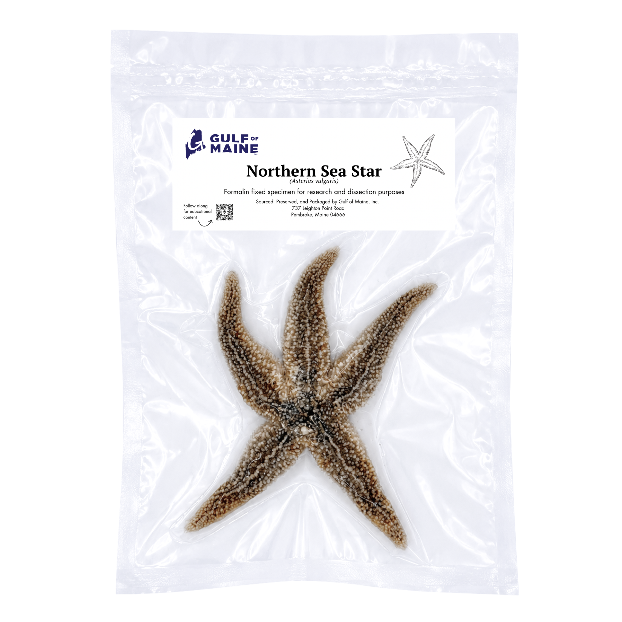
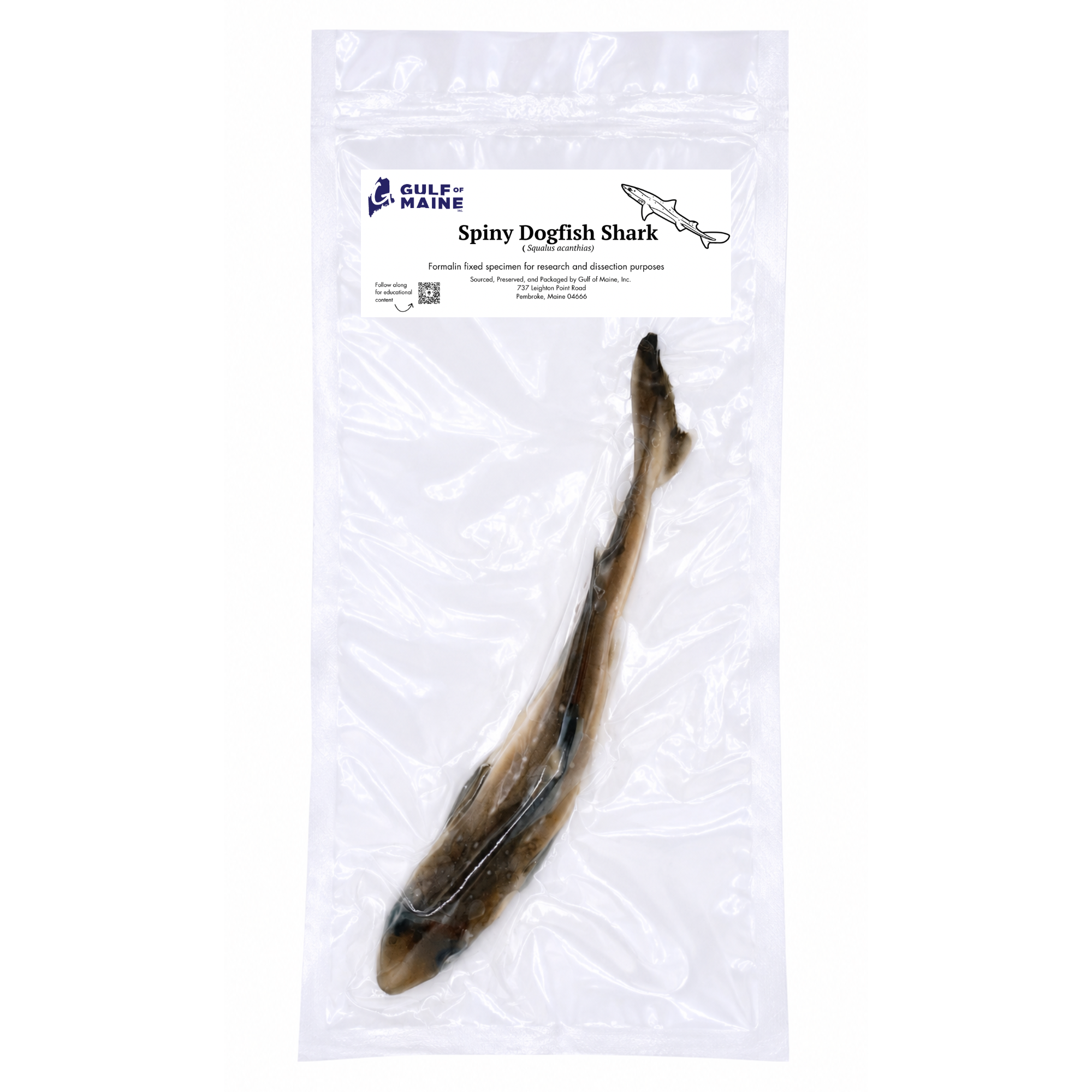
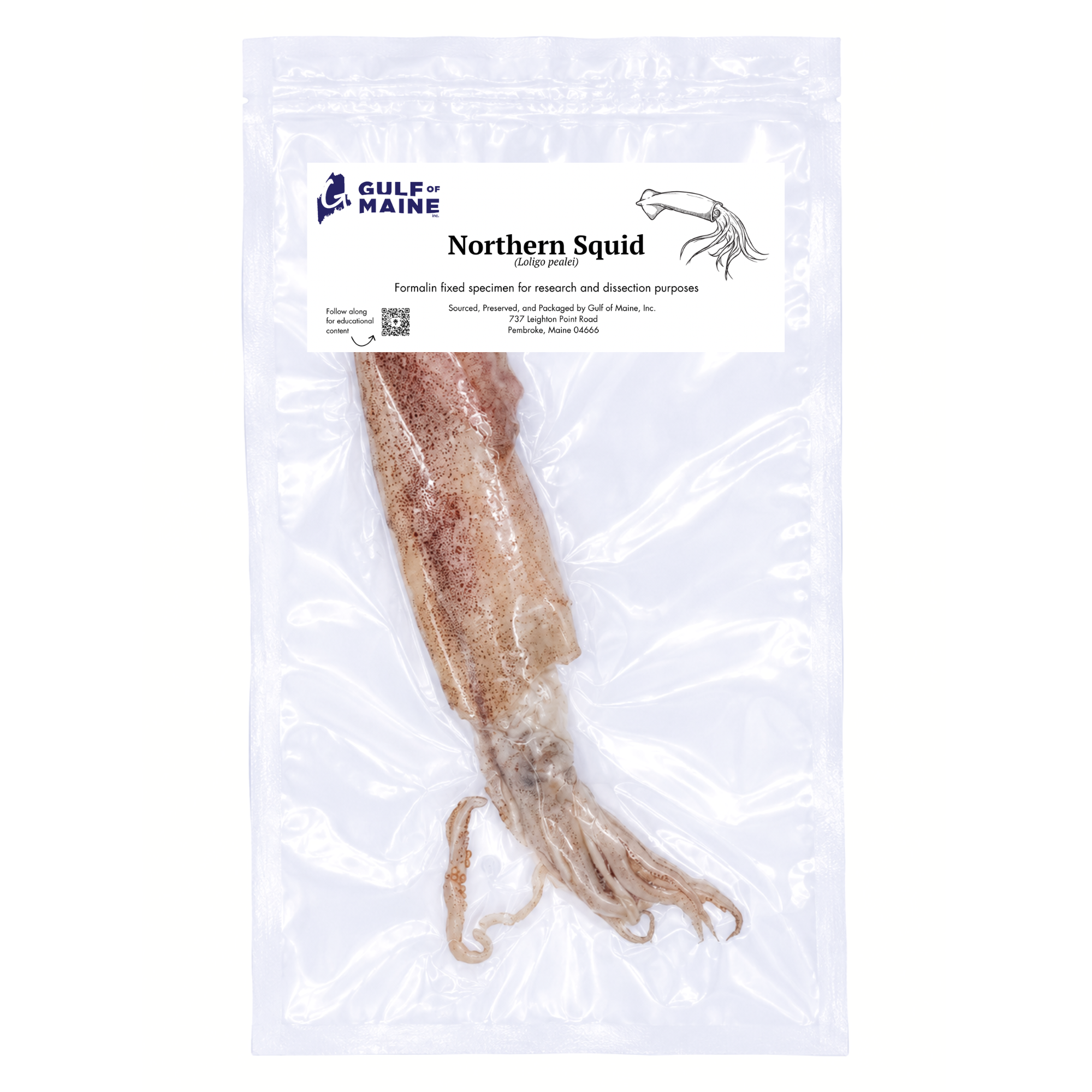
Want to learn more about some of the species featured above? Explore these trusted resources for additional information on their biology, habitat, and conservation status.

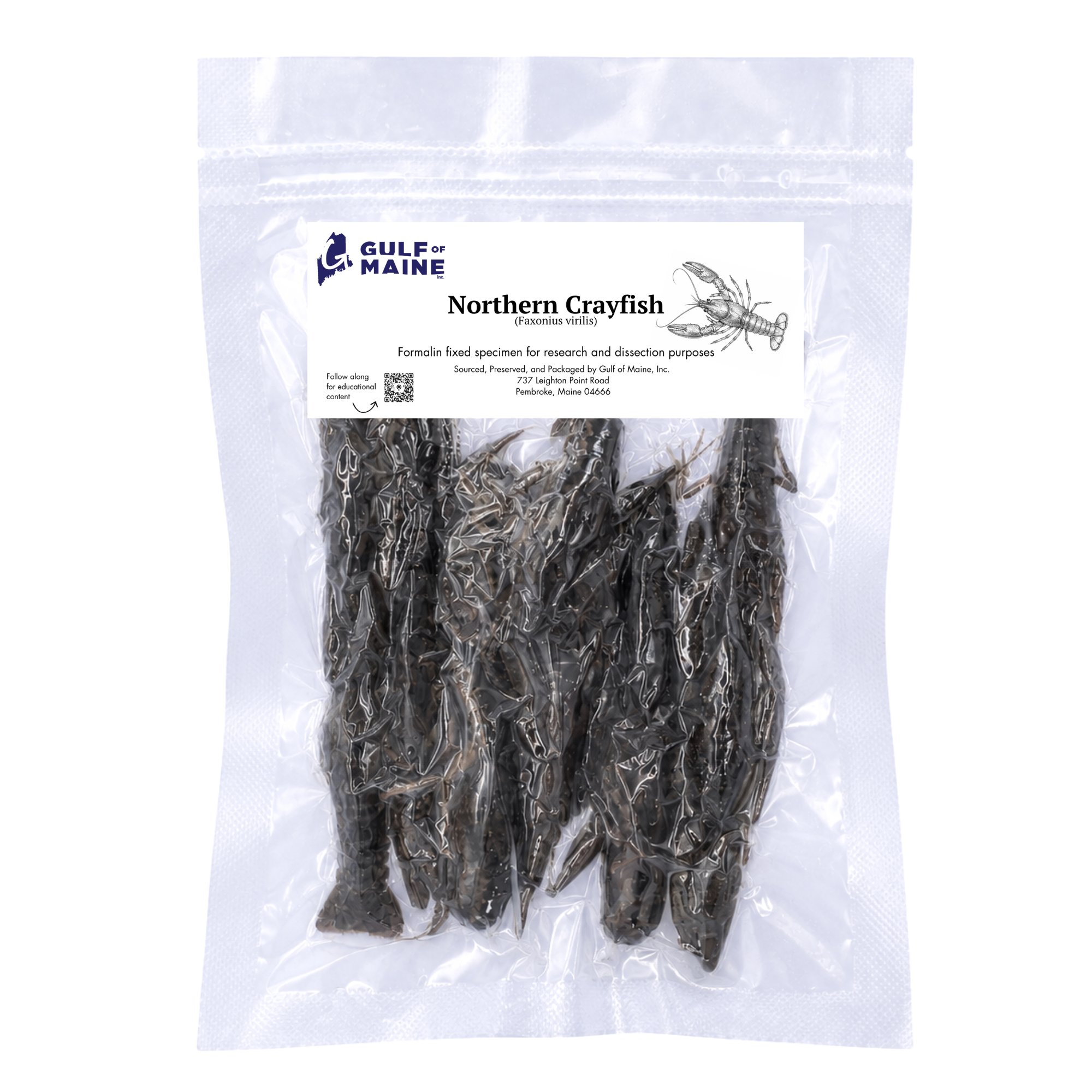
This real preserved Northern Crayfish (Faxonius virilis) provide students with a hands-on specimens for studying crustacean anatomy, arthropod biology, and comparative zoology. Professionally preserved in formalin and vacuum sealed for long-term educational use, each specimen allows detailed examination of the exoskeleton, cephalothorax, abdomen, walking legs, swimmerets, gills, compound eyes, antennae, and chelae (claws).
Ideal for biology classrooms, anatomy laboratories, homeschool science, universities, museums, and educational collections, this specimen supports lessons on arthropod anatomy, freshwater ecology, taxonomy, and comparative zoology.
Because these are natural specimens, size varies, with most measuring roughly 3–6 inches. Each specimen is professionally prepared, individually vacuum sealed, and ready for classroom or laboratory use.